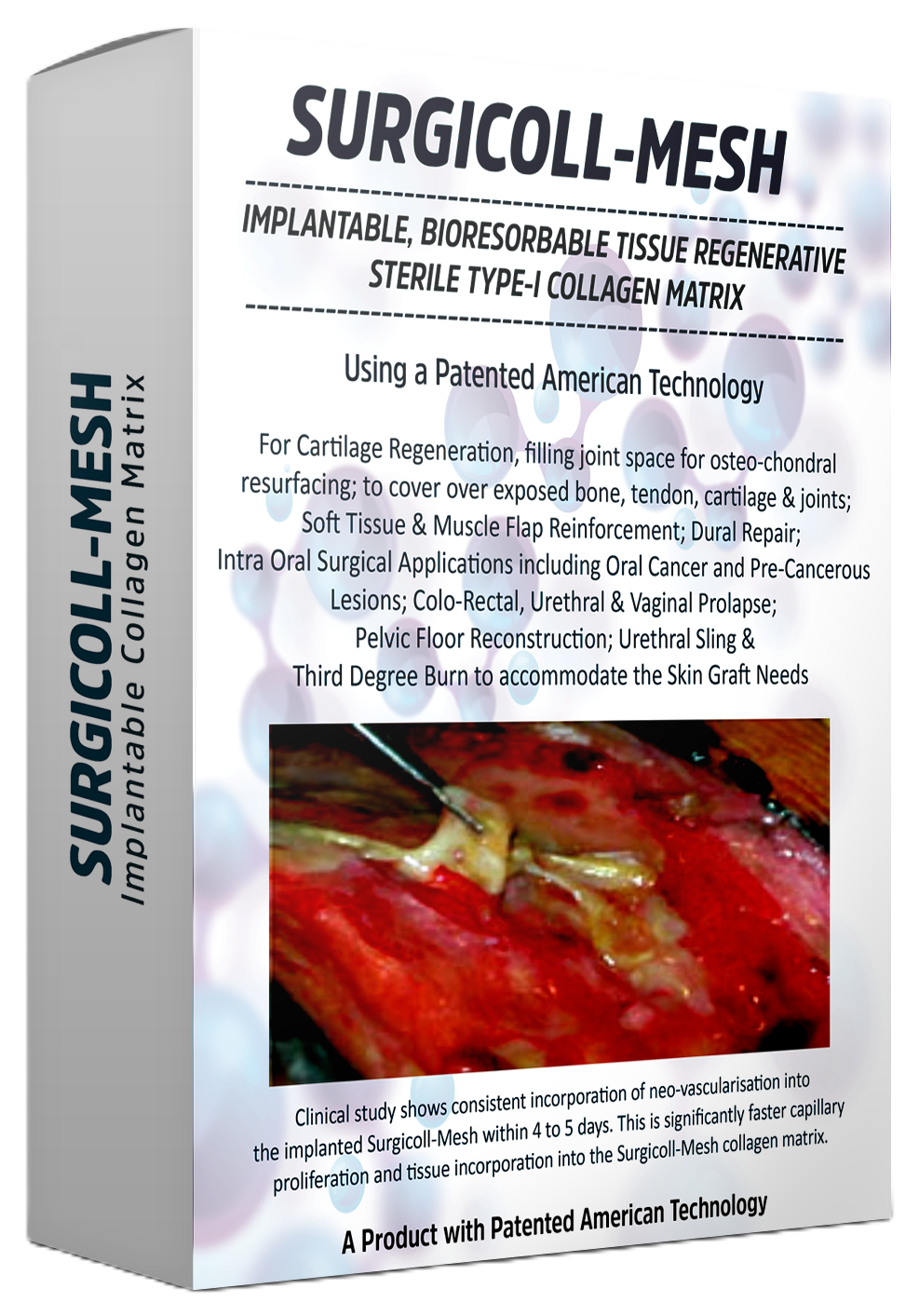
Surgicoll-Mesh®
Surgical Oncology
Implantable, bioresorbable, tissue regenerative sterile Type-I Collagen Acellular Dermal Matrix designed for reconstructive breast surgery, oral cancer and precancerous lesion repair.
Surgical Oncology
Implantable, bioresorbable, tissue regenerative sterile Type-I Collagen Acellular Dermal Matrix designed for reconstructive breast surgery, oral cancer and precancerous lesion repair.
Oral cancer can develop at multiple sites within the oral cavity and oropharynx. The most frequently involved locations include:
Pre-cancerous lesions such as leukoplakia (white patches) and erythroplakia (red patches) often precede malignant transformation and are critical to identify during routine dental or clinical examinations.
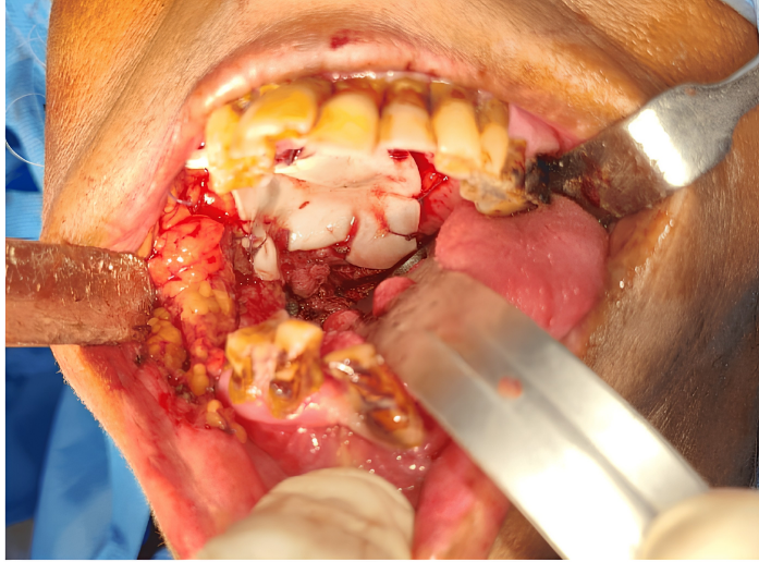
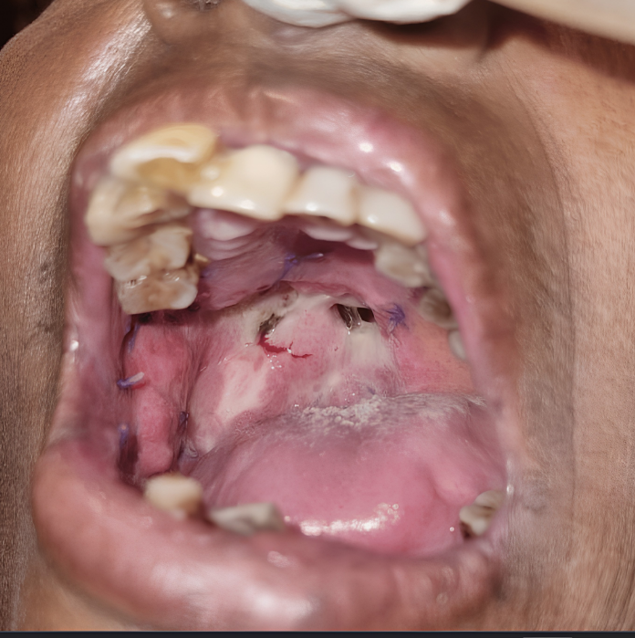
Phase 1 double-blind randomized controlled clinical trial (CTRI-REF/2023/12/076231). 20 patients divided into two equal groups.
Parameters assessed at 2 weeks: epithelialization, granulation tissue formation, and wound contraction. Independent sample t-test conducted at 95% confidence interval.
SurgiColl-Mesh® has an average porosity of 20 microns (vs. >800 microns in conventional membranes), yielding better cell infiltration and regenerative factor availability. No adverse reactions occurred in either group.
Surgical excision remains the cornerstone of oral cancer treatment. Following tumour removal, reconstruction of the defect site is essential for restoring form and function.
Bioresorbable collagen matrices such as Surgicoll-Mesh® (Sheet Form) represent an innovative approach to oral cancer excision repair. Acting as a scaffold that is naturally and progressively integrated, remodelled, and eventually replaced by functional host tissue, they offer clear advantages over synthetic alternatives in supporting healing after oncological surgery.
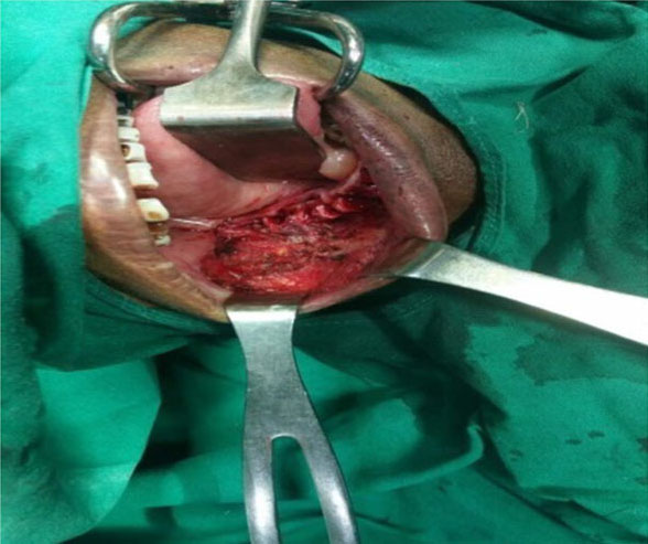
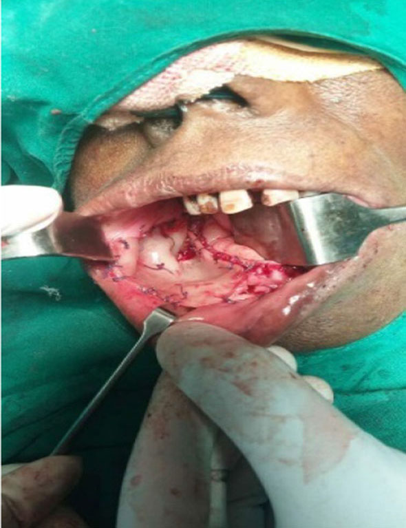
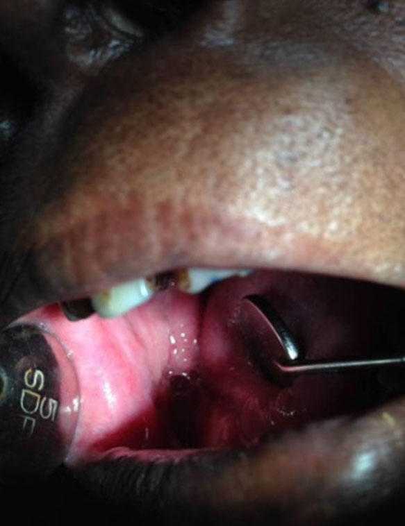
A clinical study evaluating the effectiveness of bovine-derived collagen (Surgicoll-Mesh®, 0.5 mm) in surgical oral mucosal defects. Thirty-two patients (8 female, 24 male, ages 18–65) who underwent surgery for benign, premalignant, and malignant oral cavity lesions were enrolled from 2012–2018. Cases included leukoplakia, oral submucous fibrosis (OSMF), mucocele, fibromas, and malignant lesions.
The membrane was soaked in normal saline for a minimum of 5 minutes, adapted to the mucosal defect, trimmed with scissors, and secured with 3-0 Vicryl sutures. Patients were evaluated at Day 3, 7, 30, and 3 months using the Bessho et al. scoring criteria.
| Parameter | Good | Fair | Poor |
|---|---|---|---|
| Haemostasis | 81.25% | 18.75% | 0% |
| Pain Relief | 68.75% | 31.25% | 0% |
| Granulation Tissue | 87.5% | 12.5% | 0% |
| Epithelialisation | 93.75% | 6.25% | 0% |
| Contracture Control | 62.5% | 25% | 12.5% |
| Overall Effectiveness | 81.25% | 18.75% | 0% |
Within 1 week, 75% of patients reverted back to normal eating habits. No allergic reactions were reported in any patient.
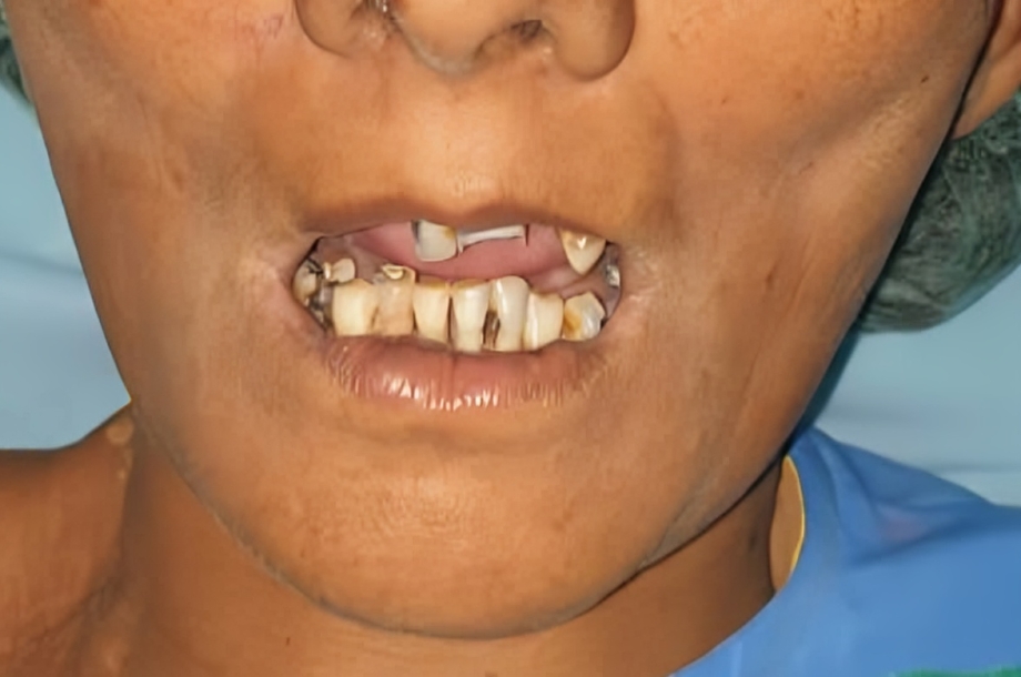
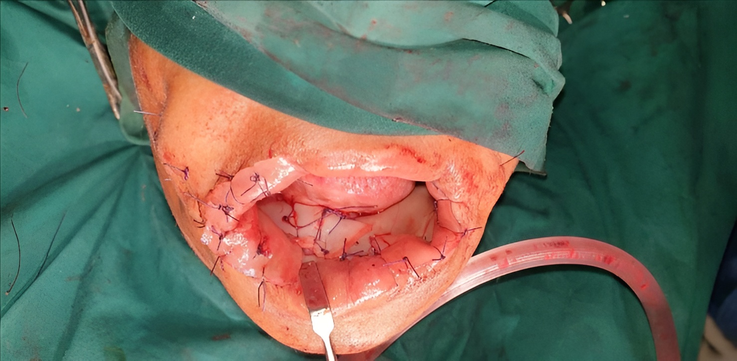
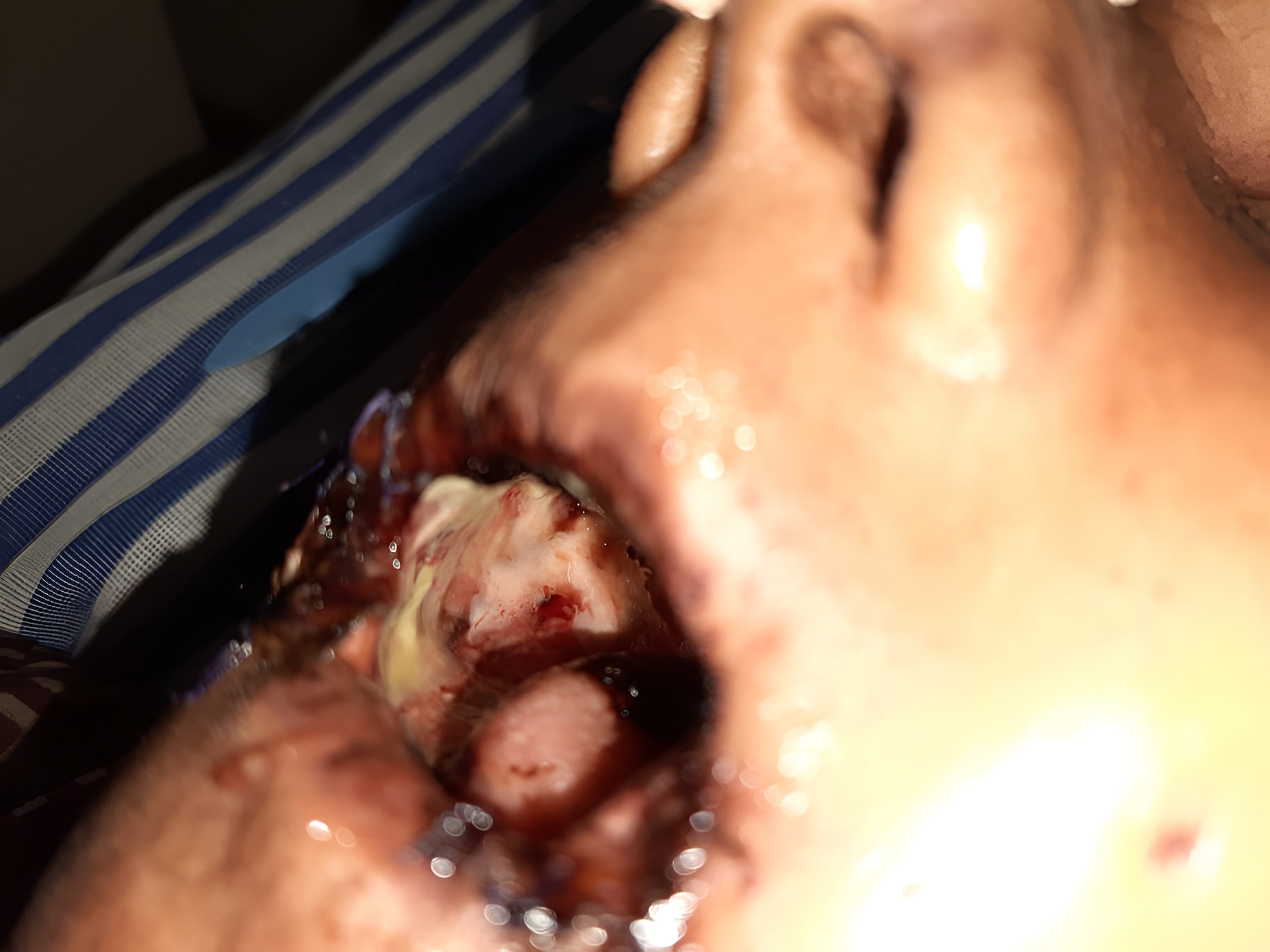
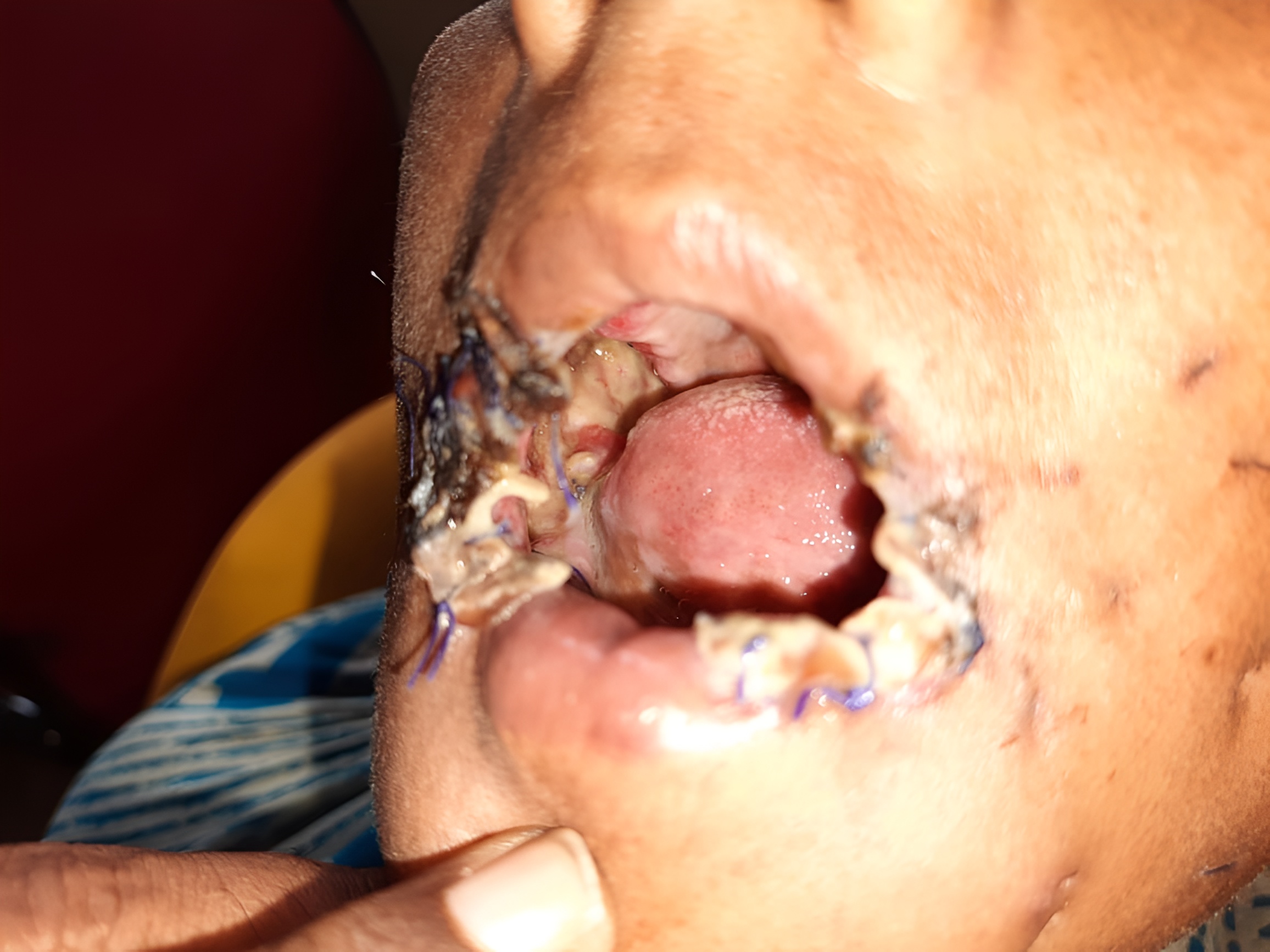
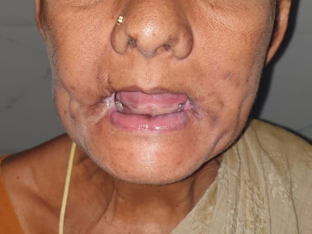
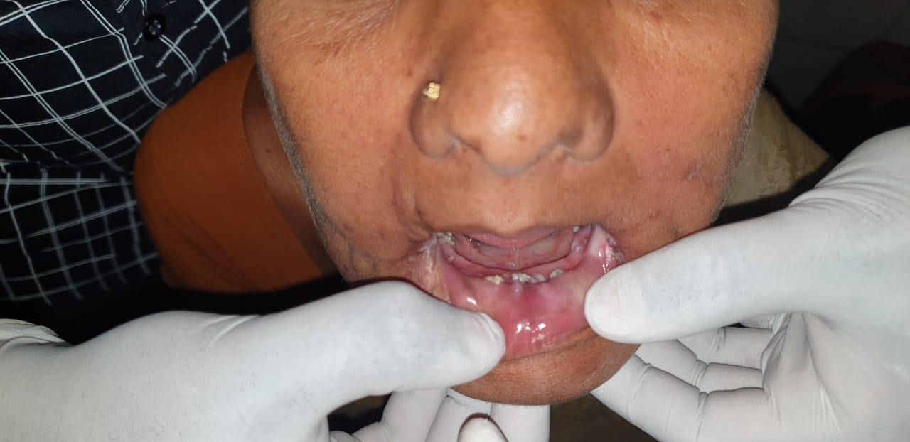
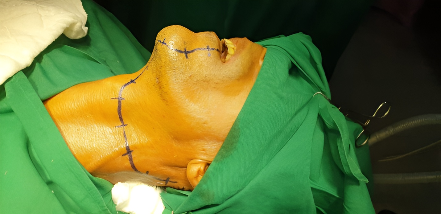
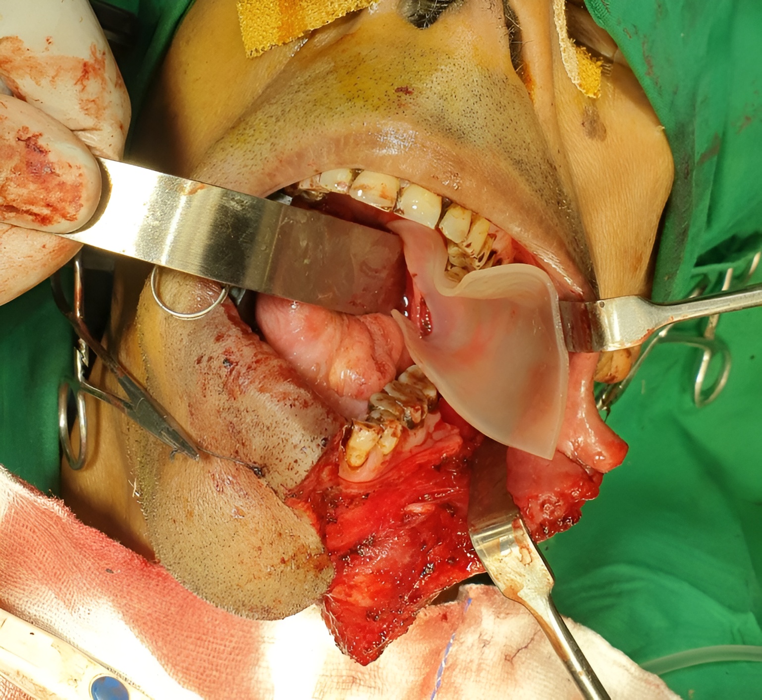
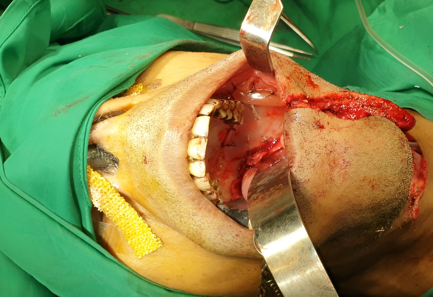
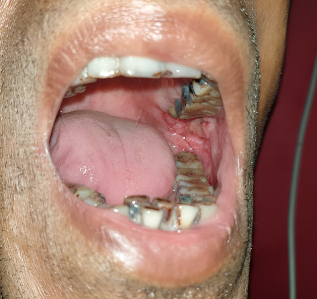
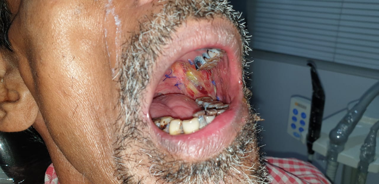
Case 2 & 3
Courtesy of Dr. Pradeep, Oral Oncologist, Nagercoil
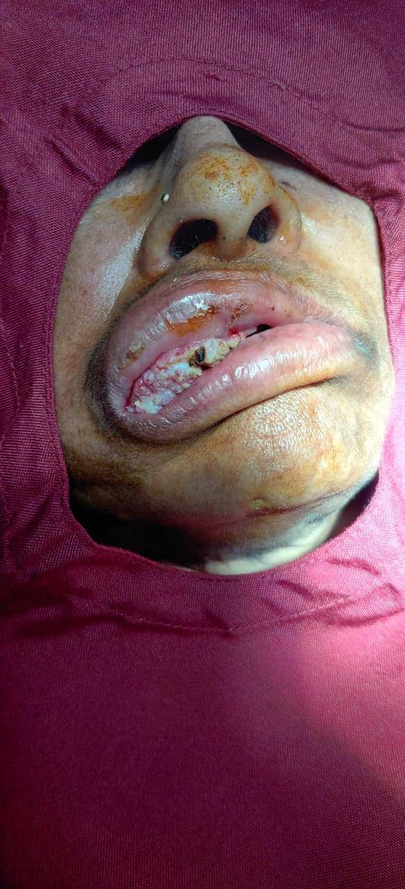
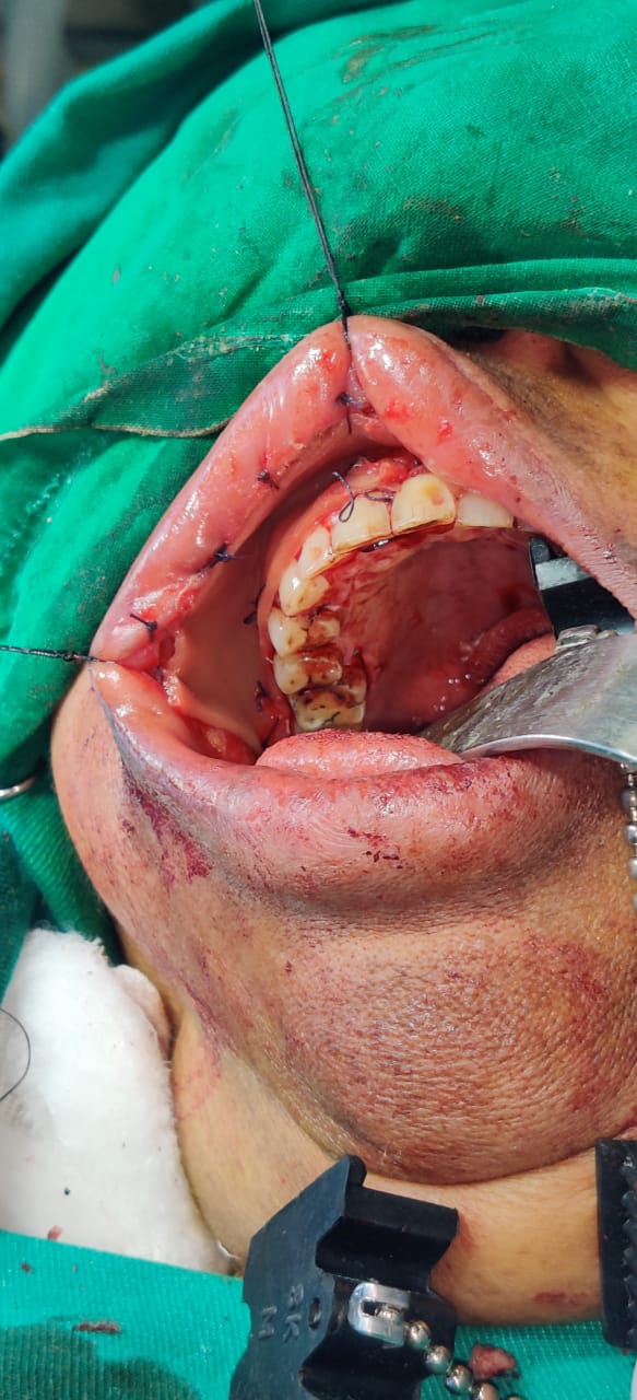
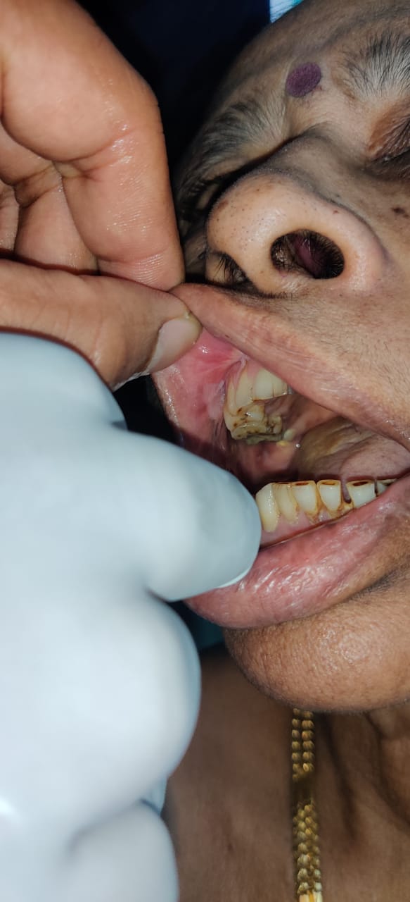
Courtesy of Dr. Mohammed Zuhaib MDS (OMFS), MFD RCSI (Ireland), MFDS RCPS (Glasgow), Fellow Oral Oncosurgery
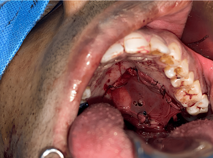
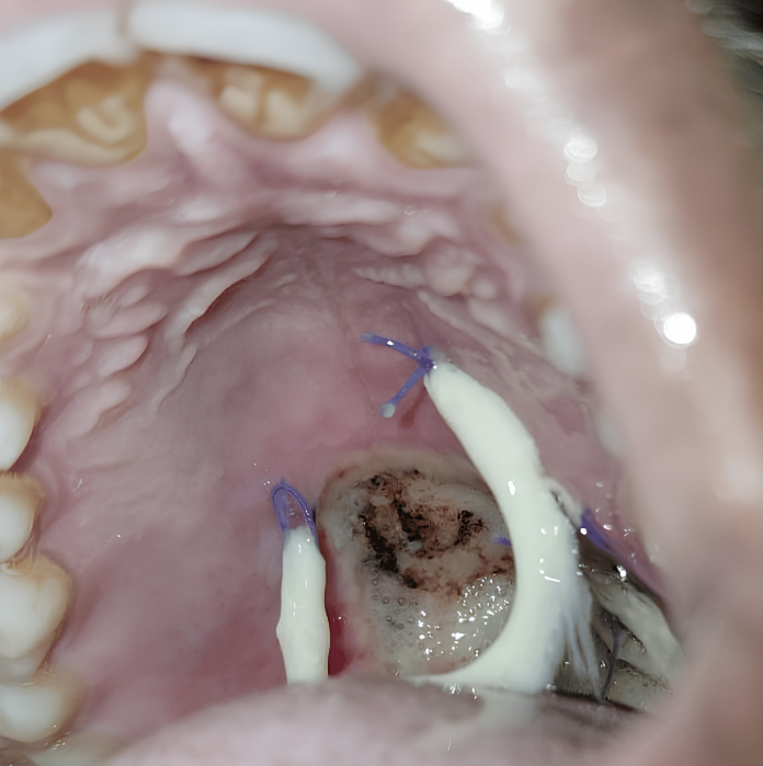
See the clinical application of Surgicoll-Mesh® in the management of post-surgical mucosal defects following oral cancer excision. The membrane becomes soft and supple when hydrated with normal saline, mimicking native tissue.
The collagen membrane is secured with 3-0 Vicryl sutures and quilted to accommodate wound drainage. Patients typically return to normal oral feeding from the 3rd post-operative day, with the membrane undergoing resorption by day 7 revealing healthy epithelial tissue underneath.
Breast cancer reconstruction (also called breast reconstruction surgery) is a surgical procedure used to rebuild the shape, appearance, and symmetry of the breast after breast cancer treatment, usually after a mastectomy (removal of the breast) or lumpectomy (partial removal of breast tissue).
Helps restore the natural contour and structure of the breast.
Improves balance and appearance after treatment.
Supports physical and emotional healing after cancer care.
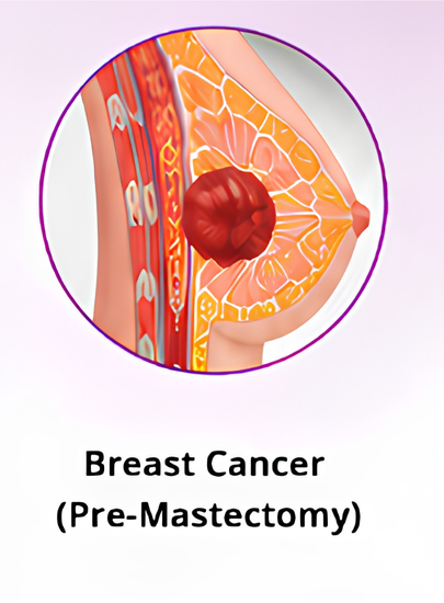
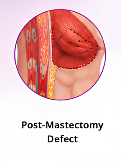
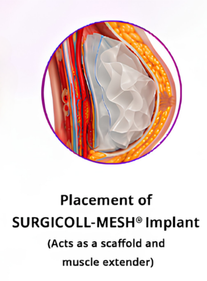
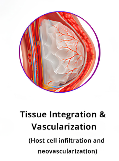
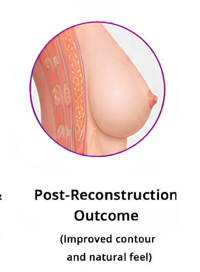
SURGICOLL-MESH® bridges the gap left after tumor excision. Once placed, the bioresorbable collagen matrix acts as a temporary scaffold that extends the pectoralis muscle, giving surgeons a stable structure for implant-based reconstruction. Over the following weeks, the patient's own cells migrate into the matrix, new blood vessels form, and the scaffold is gradually resorbed and replaced by natural tissue—restoring breast contour with a result that looks and feels like the patient's own.
Regenerative High Purity Type I Collagen Scaffold for Breast Cancer Excision Repair and Reconstruction
Krishnappa R, Narayan N, Narasimhamurthy A, Deep SS. Cureus. 2026 Mar 21;18(3):e105608. doi: 10.7759/cureus.105608. PMID: 41868445; PMCID: PMC13005701.
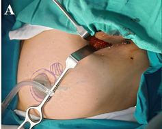
Skin marking of the breast tumour with concurrent axillary dissection in progress.
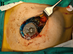
Post-excision defect in the upper outer quadrant demonstrating the residual cavity after tumour resection.
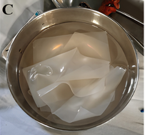
Hydration and preparation of the high-purity type I collagen scaffold prior to implantation.
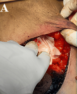
Placement of the high-purity type I collagen scaffold into the excision cavity to restore volume.
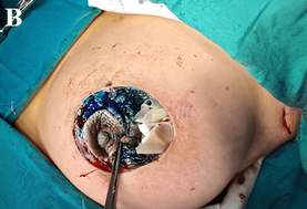
Scaffold fixation secured with absorbable sutures to surrounding breast parenchyma.
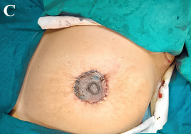
Completed layered skin closure. following scaffold implantation.
Breast-conserving surgery (BCS) causes volume displacement defects, asymmetry, and contour deformities that significantly lower post-operative patient satisfaction scores. Standard tissue substitutes carry donor site morbidity.
Implantation of the HPTC scaffold immediately following excision provides stable volume restoration, excellent early structural retention, and clear radiologic integration visible on imaging follow-up.
Demonstrated to be a completely safe and biologically active matrix option, showing high cosmetic satisfaction scores with minimal complications, zero allergic reactions, and no tissue rejection across all study patients.
Review or download comprehensive specifications, product literature, and peer-reviewed publications.
Choose from our wide range of standard sizes (in cm). Custom sizes available on request.